Comprehensive Craniometry for Sagittal Synostosis
Journal
Neurosurgical Focus
READ ONLINE
Authors
Jason A Ramsey 1 2, Phillip M Stevens 3 4, Brittany Coats 5, Timothy J Dixon 5, Sara C Chaker 6, Christopher M Bonfield 2, Michael S Golinko 6 7
- Hanger Clinic, Nashville, Tennessee.
- Departments of Neurological Surgery and.
- Hanger Institute for Clinical Research and Education, Austin, Texas.
- Departments of Physical Medicine and Rehabilitation and.
- Mechanical Engineering, University of Utah, Salt Lake City, Utah; and.
- Plastic Surgery, Vanderbilt University Medical Center, Nashville, Tennessee.
- Division of Pediatric Plastic Surgery, Cleft and Craniofacial Program, Monroe Carell Jr. Children’s Hospital at Vanderbilt, Nashville, Tennessee.
Background
Sagittal synostosis is the most common type of craniosynostosis, accounting for approximately 50% of craniosynostosis cases and results in skull deformity with distinctive characteristics. These include occipital narrowing, parietal narrowing, anteriorly shifted vertex with parietal depression, and exaggerated frontal bossing. The cranial deformities associated with craniosynostosis are complex, three-dimensional presentations that have historically been poorly captured by existing cranial indices. Despite its prevalence, there has not been a standard outcomes measurement until now.
Recognizing the inadequacy of existing measurement, the Hanger Institute for Clinical Research and Education and Hanger Clinic recently partnered with Vanderbilt University Medical Center and the University of Utah to develop a new standard to measure infants with sagittal craniosynostosis. These novel cranial indices aimed to better quantify the variety of presentations observed in infants with sagittal craniosynostosis.
Objective
To conceptualize, refine, and evaluate a set of novel metrics based on optical surface scanning (OSS) technology used for post-operative cranial remolding orthosis (CRO) treatment.
Design
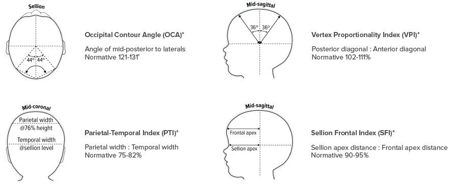
A retrospective review of the 25 most recent infants to receive CRO treatment for sagittal synostosis at a single center was completed. All treated patients underwent the endoscope-assisted craniectomy technique without barrel staving. OSS representations of each patient’s head were acquired perioperatively and at cessation of CRO treatment. A novel set of metrics were developed, comprising the occipital contour angle to assess severity of occipital narrowing; the vertex proportionality index to assess the anterior vertex relative to the depressed posterior anatomy; the parietal-temporal index to assess proximal cranial narrowing; and the sellion-frontal index as a measure of frontal bossing. The pre- and post-treatment results for all indices were compared against each other and against a control group of 33 nonsynostotic infants with grossly normal head shapes.
Results
Initial treatment group means for all 4 indices demonstrated significant variance against both the final treatment group means and the control group means.
No statistically significant differences were observed in the group means for occipital contour angle, parietal-temporal index, and sellion-frontal index between the post-treatment and control cohorts, which was suggestive of mean correction to normative levels for these morphological considerations. Despite an appreciable mean correction of parietal depression in the final treatment group, the mean vertex proportionality index values remained statistically different from the control group.
Conclusion
Sagittal synostosis is characterized by several characteristic deviations from normal. These differences are effectively improved by endoscope-assisted craniectomy with CRO intervention.
- Head shape abnormalities differ between patients, and the individual subject can present normatively for some deformational categories.
- A multimetric approach is essential to quantify initial presentation and subsequent outcome.
- OSS-enabled craniometry may facilitate more patient-centric management of this complex deformity, identifying features with the greatest deviation from normative standards and enabling creation of discrete treatment plans with respect to the focus and length of postoperative helmeting.
- These new cranial indices have been integrated into the Craniometrics Reporting Tool, allowing an unprecedented level of measuring and tracking cranial presentations and improvements.
- Hanger Clinicians can immediately apply the new Craniometrics Reporting Tool to their orthotic clinical practice, allowing them to deliver exceptional care and ultimately improve patient outcomes and satisfaction.
More Published Research
This publication is just one of the studies conducted by the Hanger Institute for Clinical Research and Education, in collaboration with leading researchers, clinical, and academic institutions.
Latest Updates
Subscribe to stay up-to-date on our latest posts.