Research Review: Quantifying Orthotic Correction of Trigonocephaly Using Optical Surface Scanning
Journal
The Journal of Craniofacial Surgery
Read Online
Authors
Jason A. Ramsey, MSPO1, Phillip M. Stevens, MEd2, Shane R. Wurdeman, PhD2, and Christopher M. Bonfield, MD3
- Hanger Clinic, Nashville, TN;
- Department of Clinical and Scientific Affairs, Hanger Clinic, Austin, TX;
- Department of Neurological Surgery, Vanderbilt University Medical Center, Nashville, TN
Objective
The purpose of this preliminary study was to evaluate a method for safely and repeatedly measuring frontal angle (FA) using technology available at multiple centers providing treatment with cranial remolding orthoses.
Technique
FIGURE 3. FA30 frontal angle measurement technique to quantify trigonocephaly and correction.
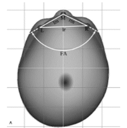
Figure 3A) Triangular relationship measured at the height of the cranium’s maximum circumference. The apex is located at the midpoint of the forehead surface (M) where the landmarks (L) and (R) are located on the forehead 30 degrees to the left and right of midline. The angle of the resultant triangle is the FA30 frontal angle measurement.
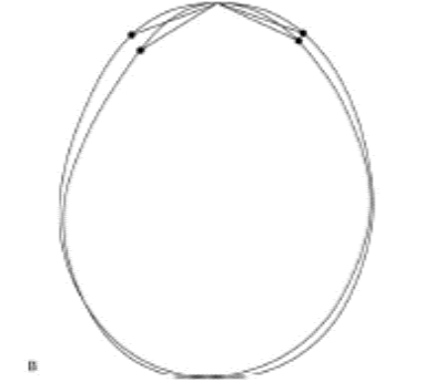
Figure 3B) Treatment subject’s superimposed initial and corrected measurement planes scaled to the same AP diameter with FA30 increasing from 125.48 to 141.08. AP, anteroposterior diameter.
Results
- All trigonocephalic subjects had initial Frontal Angle (FA) significantly lower than the control group and other cohorts (p<0.001).
- Mean FA increased from 121.5° to 138.5° (p<0.001), approaching the control group mean of 144.4°.
- Intraclass coefficient calculation showed high inter-rater reliability (ICC: 0.993, 95% CI: 0.957-0.998, p<0.001) in the technique, which was supported with Bland-Altman analyses of agreement.
- Increase in FA objectively demonstrates correction of trigonocephaly
Conclusion
- Optical surface scanning may provide a safe, accurate, and repeatable means to measure Frontal Angle (FA).
- Increase in FA30 demonstrates correction of trigonocephaly.
- The novel optical scanning technology does not subject children to radiation like traditional measures.
- The method presented enables expeditious reporting of treatment progress to the infant’s surgeon and parents, and has potential for use in optimizing treatment outcomes at multiple centers.
Introducing the CARE Network
Hanger Clinic’s cranial orthotists are equipped with advanced knowledge and unique optical scanning technology that can provide reliable, accurate measurement of the correction of trigonocephaly, including this novel scanning technique developed by CARE Network clinician Jason Ramsey.
Latest Updates
Subscribe to stay up-to-date on our latest posts.